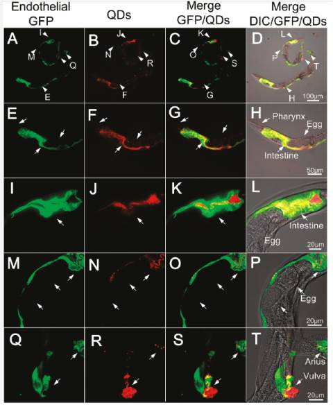
1) Establish systematic research method on biological effects of nanomaterials
We integrate optical imaging, synchrotron radiation based elemental imaging, cell biological and toxicological techniques to study the in situ metabolism and degradation of QDs in the alimentary system of Caenorhabditis elegans (C. elegans), as well as the long-term toxicity of QDs on reproduction of C. elegans. Our work provides the first example of systematical evaluation of the physiological behavior and acute/chronic toxicity of QDs by utilizing C. elegans as a model organism and will provide new systematic methods to evaluate the potential risks of nanomaterials. (Nano Letters, 2011)

(Nano Letters, 2011)
2) Establish in situ analysis method to study the nanoparticles in biological tissues
In combination of synchronization radiation and toxicological techniques, we established the two-dimensional distribution and chemical form transformation analysis methods to study the nanoparticles in biological tissues. We can use μ–XANES and μ–XRF elemental mapping which highlight the advantage of non-invasive imaging to determine the distribution of nanomaiterials in the organisms. (Toxicol Lett, 2008; Toxicology, 2008; JRNC, 2007, etc.)
1) Establish systematic research method on biological effects of nanomaterials
We integrate optical imaging, synchrotron radiation based elemental imaging, cell biological and toxicological techniques to study the in situ metabolism and degradation of QDs in the alimentary system of Caenorhabditis elegans (C. elegans), as well as the long-term toxicity of QDs on reproduction of C. elegans. Our work provides the first example of systematical evaluation of the physiological behavior and acute/chronic toxicity of QDs by utilizing C. elegans as a model organism and will provide new systematic methods to evaluate the potential risks of nanomaterials. (Nano Letters, 2011)
(Nano Letters, 2011)
2) Establish in situ analysis method to study the nanoparticles in biological tissues
In combination of synchronization radiation and toxicological techniques, we established the two-dimensional distribution and chemical form transformation analysis methods to study the nanoparticles in biological tissues. We can use μ–XANES and μ–XRF elemental mapping which highlight the advantage of non-invasive imaging to determine the distribution of nanomaiterials in the organisms. (Toxicol Lett, 2008; Toxicology, 2008; JRNC, 2007, etc.)
 Close Page
- Text Size: A A A
 Printer Friendly