On November 13, 2023, Nature Protocols published an original methodology paper of Prof. CHEN Chunying’s group from the National Center for Nanoscience and Technology (NCNST) of the Chinese Academy of Sciences (CAS), entitled In situ label-free X-ray imaging for visualizing the localization of nanomedicines and subcellular architecture in intact single cells .
The understanding of underlying principles and mechanisms of nano-cell interactions is of great significance to accurately perceive the biological effects of nanomedicines, guiding the precise design of nanomedicines with high efficacy and negligible toxicity, eventually supporting the clinical translation. However, the complexity ofcellular structures and dynamic changes in chemical forms of nanomedicines pose great challenges for in situ label-free analysis of single cells at nanometric resolution. Synchrotron radiation-based techniques have multiple advantages of label-free, in-situ, high resolution, element-specific, quantitative analysis, and simple sample preparation. These advanced techniques are conducive to the study of the biological behavior and fate of nanomedicines in cells or tissues with native or near-native states, providing information on the spatial distribution, volume, number, density, and even the valance state of nanomedicines, as well as the morphological changes of cells or tissues.
For more than two decades, Prof. CHEN's group has been at the forefront of developing innovative analytical methods using synchrotron radiation light source facilities. They have successfully broken through the technical barriers of in situ, label-free analysis of nanomaterials in complex biological systems, paving the way for a new research paradigm for the cross-scale analysis of nanomaterials at molecular, interface, cell, tissue and in vivo levels. These groundbreaking methods have been instrumental in quantifying the interaction of nanoparticles with macromolecules (proteins, phospholipids, etc.) at the nanoparticle-biological interface and qualitatively characterizing the chemical behavior of nanomaterials within organisms. As a result, they have achieved a series of innovative research findings (Nature Nanotechnology, 2021, 16: 708; Nature Nanotechnology, 2022, 17: 993; Science Advances, 2023, 9: eadg2252; Accounts of Chemical Research, 2019, 52: 1507; Journal of the American Chemical Society, 2013, 135: 17359; Chemical Society Reviews, 2013, 42: 8266; ACS Central Science, 2022, 8: 1063; etc.). These advanced techniques are key and cutting-edge analytical tools for the study of nano-biological effects, and greatly promote the development of nanomedicines.
The Protocol paper summarized the technical details of synchrotron radiation soft and hard X-ray microscopy pioneered and established by Prof. CHEN’s group (Figure 1a), realized the in situ, label-free and 2D/3D imaging of single cellular structures and nanomedicines, as well as the chemical transformations of nanomedicines. An optimized workflow which includes cell sample preparation, hard and soft X-ray beamline selection, data acquisition and analysis was proposed for investigating the cellular localization of nanomedicines and the morphology of organelles (Figure 1b). Besides, by taking several model nanomaterials as examples, we provided a practical guideline to allow users to select proper X-ray imaging methods. The protocol can be completed by any researcher with basic biology and chemistry skills in approximately 2-5 days.
The research team proposed a systematic research strategy for investigating the subcellular structures and intracellular behaviors of nanomedicines in situ and label-free, utilizing synchrotron radiation X-ray imaging. “This research strategy not only aims to elucidate the fundamental principles and mechanisms underlying nano-cell interactions, but also serves as a potent tool for exploring the alterations in cellular components, the homeostasis of bioelements, the pharmacology and toxicology evaluation of metal-based materials. This protocol is valuable for guiding researchers in selecting appropriate X-ray imaging methods, preparing samples and processing data, holding immense potential across various disciplines, including chemical metrology, cell biology, nanomedicine and materials science, etc.” CHEN Chunying says, a Professor of NCNST and the corresponding author of the current work.
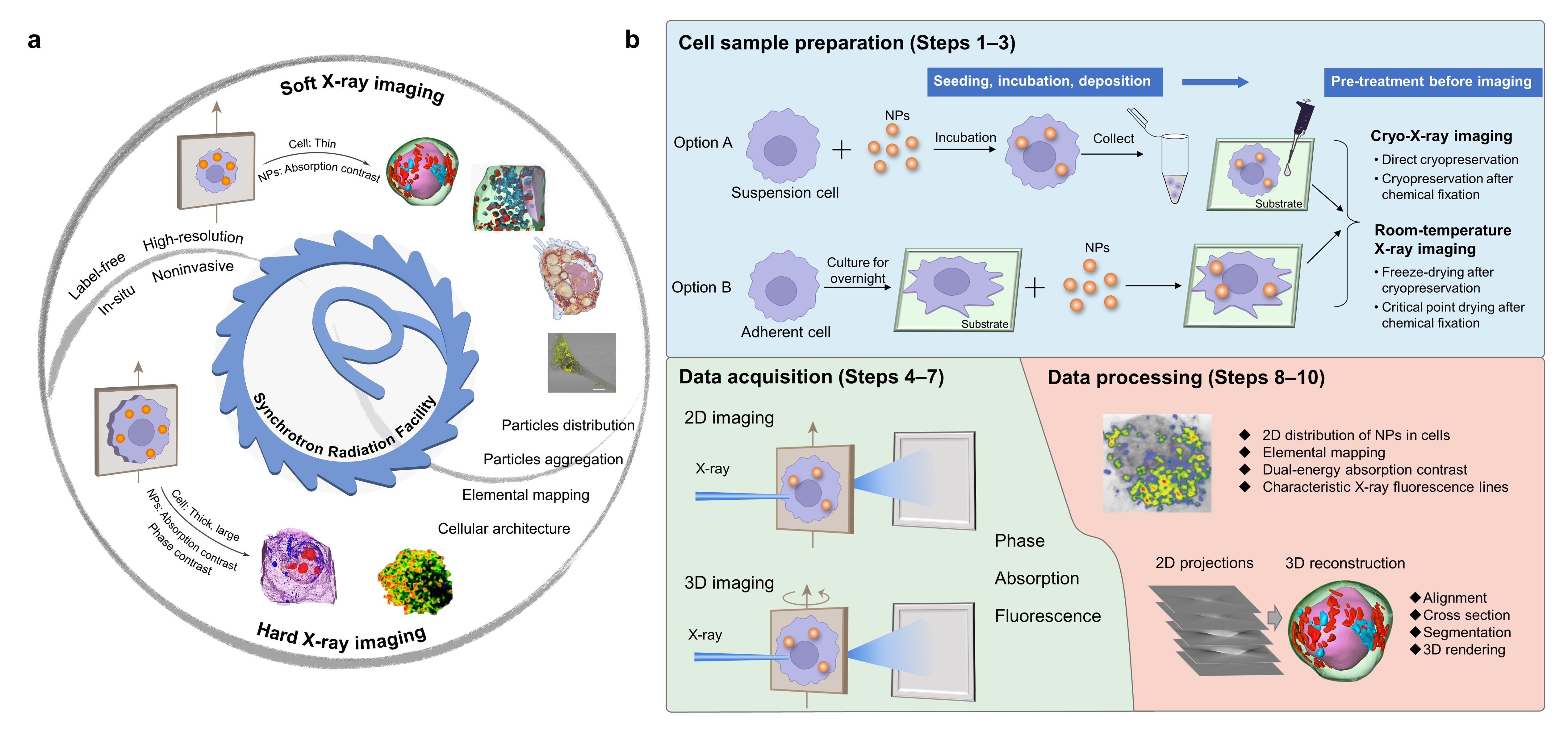
Figure 1. Soft and hard X-ray imaging of nanomedicines and subcellular architecture in intact cells based on synchrotron radiation facilities as a universal and robust platform. (Image by CHEN Chunying et al)
Dr. CAO Mingjing (NCNST) and Prof. WANG Yaling (NCNST) contributed equally to this work. The protocol was developed with the support of Prof. ZHANG Kai from Beijing Synchrotron Radiation Facility and senior engineer GUAN Yong from National Synchrotron Radiation Laboratory. This work was funded by the National Key Research and Development Program of China, the National Natural Science Foundation of China, the Strategic Priority Research Program of the Chinese Academy of Sciences (B), the National Postdoctoral Program for Innovative Talents and Synchrotron Radiation Facilities, etc.
Contact:
CHEN Chunying
National Center for Nanoscience and Technology (NCNST)
E-mail: chenchy@nanoctr.cn

