Recently, the research team led by Prof. NIE Guangjun from National Center for Nanoscience and Technology (NCNST) of the Chinese Academy of Sciences andProf. YU Jiakuo from Tsinghua Changgung Hospital Medical Center of Tsinghua University reported thatchondrocyte-mimicking nanoparticles containing a Wnt pathway inhibitor adhere to cartilage for improved treatment in osteoarthritis (OA) models. The study was published in Science Translational Medicine (2024, DOI:10.1126/scitranslmed.adh9751).
OA is a highly prevalent degenerative joint disease, characterized by local progressive articular cartilage degradation, leading to chronic pain and eventual functional failure of individual joints. However, there are currently no disease-modifying treatments available in clinical practice.One of the major challenges for pharmacotherapies in the treatment of OA is the long-term maintenance of effective drug concentrations around the chondrocytes. However, even with direct intra-articular injection, drugs are rapidly eliminated from the joint cavity through synovial capillaries and lymphatic drainage. Meanwhile, the spatially dense cartilage extracellular matrix (ECM) presents a substantial steric hindrance to the permeation of therapeutic molecules and further internalization by chondrocytes.
“An ideal drug delivery platform for the treatment of OA would be able to sufficiently permeate cartilage before being cleared, and bind to sites within the cartilage, transforming the barrier into a drug reservoir for sustained intra-cartilage delivery.” said Prof. YU Jiakuo, one of the paper’s corresponding authors.
The research team directly leveraged naturally-derived chondrocyte membrane coating as a means of bestowing cartilage drug delivery systems with enhanced binding capabilities. The engineered chondrocyte membrane-decorated poly(lactide-co-glycolic acid) (PLGA) nanoparticles (CM-NPs) retained critical chondrocyte membrane proteins that supported cell and matrix interactions in rat and human cartilage explants. These nanoparticles were retained in rat knee in vivo for at least 34 days. Simulated synovial fluid clearance studies showed that CM-NPs loaded with a Wnt pathway inhibitor (CM-NPs-Ada) delayed the catabolic metabolism of chondrocytes and cartilage explants under inflammatory conditions. In rat and canine OA models, CM-NPs-Ada effectively restored gait, attenuated peri-articular bone remodeling, and provided long-term chondroprotection against cartilage degeneration. Overall, the chondrocyte-mimicking particles offer an opportunity for the development of cartilage drug depot to improve the pharmacokinetics and efficacy of other anti-OA drugs.
“We were inspired by how chondrocytes interact with the extracellular matrix. For example, type II collagen has binding sites for α1β1, α2β1, and α10β1 integrins, whereas the fibronectin binding integrins include α3β1 and αVβ1. We wondered if we could design nanotherapeutics to mimic chondrocytes. This could allow the particles to penetrate cartilage while also adhering to cartilage fibers like chondrocytes do. In this way, we could use the body's own cartilage ECM to create a drug depot.” said NIE Guangjun, a Professor of National Center for Nanoscience and Technology (NCNST) and one of the paper’s corresponding authors.
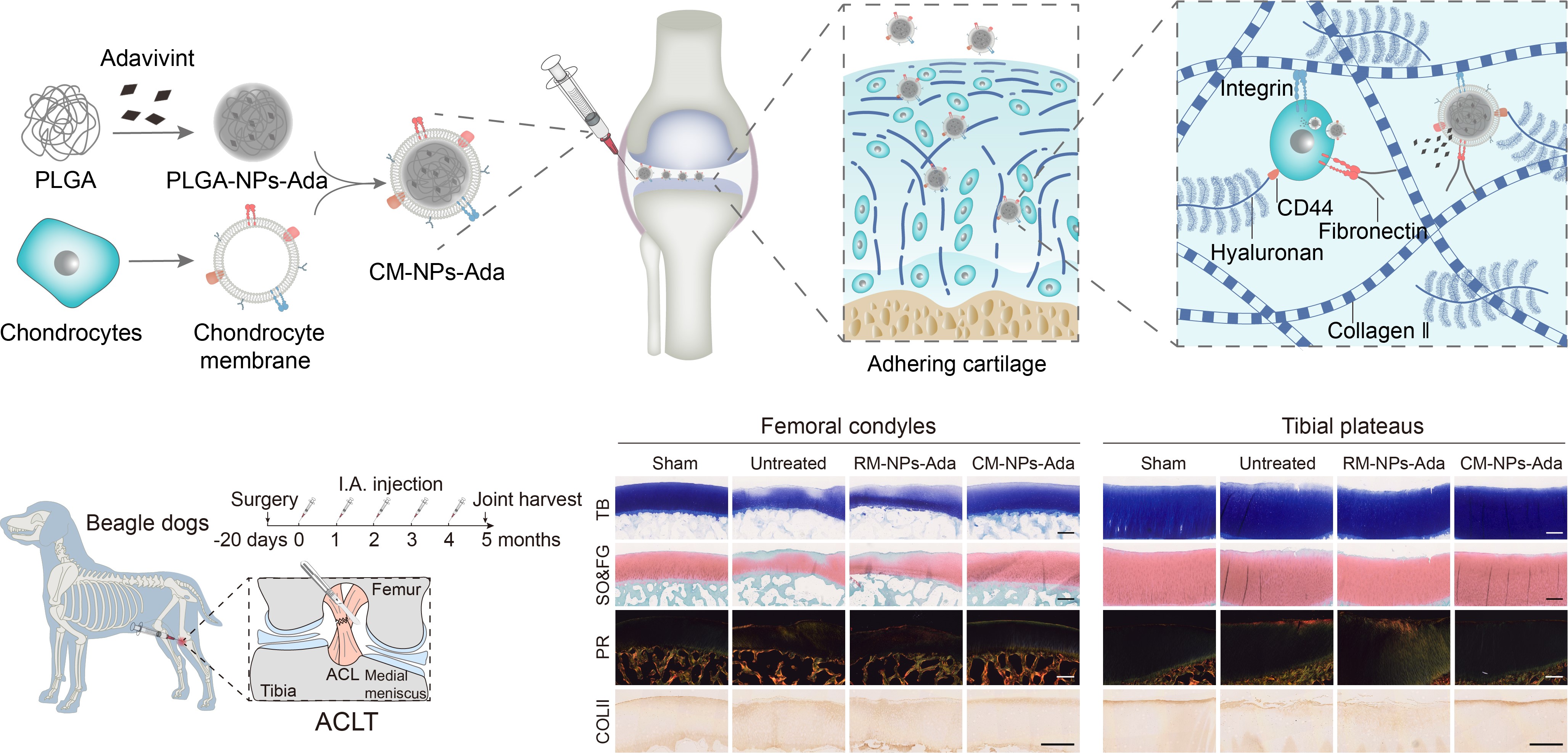
The preparation process of chondrocyte-mimicking nanotherapeutics, mechanism of action, and assessment of efficacy in large animal models (Image by NIE Guangjun et al)
Contact: NIE Guangjun
National Center for Nanoscience and Technology (NCNST)

